Femoroacetabular hip impingement (FAI)
AnatomyCausesSigns and SymptomsDiagnosisTreatmentInjections
What is femoroacetabular hip impingement (FAI)?
Femoroacetabular hip impingement (FAI) can occur for several reasons and often it is a combination of these that contribute to the diagnosis. FAI commonly occurs due to a variation in the bony anatomy and/or a labral tear of the hip joint. There are two types of anatomical variation which can lead to femoroacetabular hip impingement:
- A cam lesion – this is an excess of bone on the femoral head/neck
- A pincer lesion – this is an excess of bone on the acetabulum of the pelvis.
When one, or both, of these lesions are present, they can cause pinching or impingement of the hip joint structures (mainly the cartilage around the edge of the joint). Over time this impingement of the hip joint can cause pain, especially with prolonged periods of sitting or during and after exercise. A key finding associated with femoroacetabular hip impingement is a labral tear. The labrum is a layer of cartilage which encircles the socket of the hip joint. It is susceptible to damage when repetitive impingement takes place.
What are the symptoms of femoroacetabular hip impingement?
The symptoms of femoroacetabular hip impingement often include the following:
- A deep pain at the front of the hip/groin that can radiate to the outside of the hip and even into the buttocks. This can either be felt as a sharp pain, or a dull ache.
- Pain is increased with prolonged sitting, standing after sitting, walking and exercise, especially twisting and turning sports such as football.
- A general feeling of stiffness in the hip.
- A feeling of weakness around the hip.
- Clicking and a sensation of giving way are also sometimes reported with femoroacetabular hip impingement.
What other conditions can present like a femoroacetabular hip impingement?
- Greater trochanteric pain syndrome
- Hip osteoarthritis
- Osteitis pubis
- Iliopsoas tendinopathy/bursitis
- Proximal hamstring tendinopathy
Femoroacetabular hip impingement vs hip osteoarthritis
A femoroacetabular hip impingement is a joint related issue. It most commonly affects young people between 20 and 40 years old and is often associated with poor muscle control around the hip, stiffness of the hip and sub-optimal movements during walking, running or sport.
Hip osteoarthritis is also a joint related pathology. Osteoarthritis is a progressive, degenerative condition in which the cartilage layer of the joint (which provides friction reducing movement) becomes damaged. Osteoarthritis is a painful condition which is accompanied by joint stiffness. This it is most commonly seen in people over the age of 50 and typically advances with age.
Both femoroacetabular hip impingement and osteoarthritis of the hip are predominantly felt in the front of the hip and in the groin. Furthermore, both conditions report early morning stiffness in the hip. Both conditions can be very painful, especially with hip flexion activities but femoroacetabular hip impingement is more common in the younger population in contrast to hip osteoarthritis.
Anatomy of the hip
The hip joint is formed from by the articulation of the large ball-shaped head of the femur (long bone of the thigh) and the cup-shaped socket of the pelvis (called the acetabulum).
This large load-bearing joint is supported by a fibrocartilage ring which surrounds the acetabulum, known as the labrum. The labrum increases your joint stability by deepening the socket of the acetabulum. The hip joint is covered in articular cartilage and is bathed in a friction-reducing fluid called synovial fluid.
What is femoral acetabular hip impingement (FAI)?

Femoral acetabular hip impingement can occur as a result of subtle structural variances within the bony anatomy of the hip. When these variances are present, the acetabulum and the femoral head and neck can come into contact with each other, causing pain and impingement (Hendry et al., 2013).
There are two key structural (bony) changes associated with femoral acetabular hip impingement. These are as follows;
- Cam lesions - this refers to an excess of bone located on the head/neck of the femur. As the hip moves this excess of bone can impact with the acetabulum, causing a bony pinch or ‘impingement’.
- Pincer lesions - this refers to an excess of bone located on the rim of the acetabulum. Once again, as the hip moves, this bony protuberance catches on the femoral head/neck causing direct injury to the femur. Hip joint pathology can also occur at the opposite side of the joint due to a lever-action caused by the pincer lesion.
- It is not uncommon for some patients to experience both Cam and Pincer lesions at the same time (Cheatham et al., 2016).
Femoral acetabular impingement is a common cause of hip pain in both young and middle-aged patients and has been associated with the development of secondary hip osteoarthritis (Park et al., 2012).
Load-bearing joints such as the hip and knee are the most commonly affected by osteoarthritis [cornerstone hip article]. Current research has shown that articular cartilage requires good quality joint movement to maintain its health. Inappropriate movement patterns or sustained suboptimal loading can cause the articular cartilage to become thinned and compromised (Clarke et al., 2003). The process results in the degenerative process known as osteoarthritis.
Fortunately, research has also demonstrated that maintaining good flexibility and strength around the joint will help to slow the disease progression associated with osteoarthritis (Hunter et al, 2009).
Cheatham et al., (2016) states that the development of femoral acetabular impingement is mainly congenital. However, there are a number of risk factors associated with femoral acetabular impingement (FAI). These include;
- Hip joint hypomobility (reduced range of motion)
- Muscular stiffness/tightness in surrounding hip musculature
- Suboptimal gait pattern (differences in the way you walk have been observed in patients with FAI)
- Hip muscle weakness is regularly noted in patients with FAI (46% of FAI patients have been shown to exhibit weakness in the lateral gluteal muscles, used to stabilise the hip joint)
- Changes in the position of the pelvis have also been observed in patients with FAI
- The chance of FAI is increased after hip fracture
How do you know if you have a femoral acetabular impingement (FAI)?

Research conducted by Hendry et al (2013) and Cheatham et al (2016) describes common symptoms associated with FAI as including the following;
Signs and symptoms of FAI
- Pain can be described as either sharp or a deep aching pain located in the anterior (front) of the groin
- Pain may also be experienced on the outside aspect of the hip and into the buttocks
- A sensation of stiffness or reduced range of movement at the hip
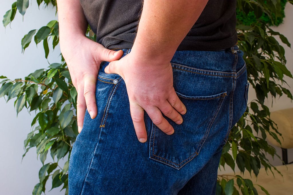
- Patients often cup their hand around the front and side of the hip when discussing the location of symptoms. This is known as the ‘C sign’ and is indicative of hip joint pathology (see above image)
- Pain which worsens with prolonged periods of sitting standing and walking (research is showed this to be present in over 96% of FAI patients)
- Hip pain is most prevalent in tasks requiring hip flexion, adduction and internal rotation such as crossing legs or hip stretches such as the images below
- Pain during sporting activities requiring rotating or pivoting on the hip such as football
- Many patients experience clicking and a sensation of giving way when suffering from FAI
- Hip muscle weakness has been shown to be present in many cases of FAI. This can result in worsening of symptoms due to loss of control of the joint during movement


How do we diagnose FAI?
To ensure the treatment you receive is effective, it is essential that an accurate diagnosis of FAI is made as early as possible.
It is not uncommon that FAI is misdiagnosed, as there are many conditions that can present quite similarly. It is recommended that you seek professional help, from one of our physiotherapists, if you feel you may be suffering from this condition.
There is a multitude of other pathologies that can masquerade as a femoral acetabular impingement that need to be excluded in order to formulate a diagnosis of FAI. These include;
- Hip osteoarthritis
- Labral tears
- Psoas tendinopathy/bursitis
- Discogenic lower back pain
- Groin strain
- Hernia
An accurate diagnosis requires a detailed clinical examination and diagnostic imaging. These will be described below:
Clinical examination
A clinical examination is used to inform the physiotherapist of the factors involved in the development of your symptoms. It is used to develop a hypothesis as to why you are experiencing your pain and to provide a treatment plan to address these issues.
A clinical interview is used to understand your situation. You will be asked questions including how and why your pain started, what aggravates and eases your symptoms and how long have you been experiencing pain. A full medical history will also be taken.
A physical examination is then completed and includes hip joint range of movement testing, strength and flexibility of the surrounding musculature, palpation (feeling) of different structures of the hip. A series of functional testing is also completed which often include squatting, single leg balance, gait analysis and running analysis (if you are a runner).
This process provides a wealth of information about what is causing your symptoms and can diagnose femoral acetabular impingement. However, imaging is required to determine why you have this condition, for example, do you have a cam and/or pincer or a tear of the labrum (cartilage around the hip socket), known as a labral tear.
Diagnostic imaging
Diagnostic imaging is essential for confirming the presence of femoral acetabular impingement (Cheatham et al., 2016).
X-ray

If your clinician suspects femoral acetabular impingement you may be referred for an initial x-ray. This is commonly used to assess bone and joint structure and is capable of assessing the shape of the bones of the hip joint as well as the quality of the joint space and presence of degenerative change.
Magnetic resonance imaging (MRI)

MRI imaging is highly accurate at assessing both bone and soft tissue pathologies. Research has shown MRI to be a gold standard diagnostic tool for assessing femoral acetabular impingement. An MRI can take between 30 minutes and an hour and involves you lying, motionless in the metal cylinder. An MRI takes a series of images of your hip to create a detailed 3D image of your hip architecture.
Diagnostic ultrasound imaging
Diagnostic ultrasound imaging is able to visualise the outer edge of the hip joint and the inflammation associated with femoral acetabular impingement. It also has the added benefit of being able to visualise the hip while moving and is therefore of great benefit when assessing for pain during certain movements.
Diagnostic musculoskeletal ultrasound imaging is not able to fully assess for hip joint pathology due to the depth of the joint, however, it is an excellent imaging tool for assessing for soft tissue hip pathology (Cheatham et al., 2016).
Complete has a highly skilled team of clinicians who are qualified as both physiotherapists and musculoskeletal sonographers. During your assessment, your clinician will use both clinical assessment and diagnostic imaging to help diagnose the cause of your hip pain.
If you would like more information or would like to book an appointment please contact us on 0207 482 3875 or email injections@complete-physio.co.uk.
How do we treat femoral acetabular impingement syndrome?

Most patients with FAI are not able to fully assess for hip joint pathology due to the depth of the joint. However, it is an excellent imaging tool for assessing for soft tissue hip pathology (Cheatham et al., 2016). Femoral acetabular impingement normally responds very well to physiotherapy.
- A personalised, progressive rehabilitation program including stretching and strengthening exercises for the hip muscles.
- Advice on activity modification to minimise the chance of irritating your symptoms.
- Movement re-education - this may include assessing and modifying how to squat or lunge correctly.
- Soft tissue release techniques may also be used to reduce pain and increase flexibility around the hip.
Here are a few top tips you may wish to try yourself:
- Avoid aggravating your symptoms - prolonged periods of sitting and crossing your legs are often associated with irritating a hip with femoral acetabular impingement. Try to avoid these if possible. If you are a runner this may mean significantly reducing your running. You may need to rest completely from running for a short period of time, stopping running for non-weight bearing exercises such as cross trainer can help.
- Try gently stretching your hip muscles particularly the hip flexors (see below)
- Pilates exercises such as the bridge can help increase the strength of your hip muscles (see below image).


What if conservative treatment does not work for femoral acetabular impingement?
If physiotherapy treatment has been ineffective at reducing pain and symptoms and the diagnosis has been confirmed on imaging then an ultrasound-guided injection may be appropriate for you.
Patients who respond particularly well to the inclusion of corticosteroid injections normally have pain that:
- wakes you at night and affects your ability to go to sleep
- is limiting your ability to complete daily living tasks including work and leisure activities.
- affects your ability to partake in physiotherapy rehabilitation
Ultrasound-guided corticosteroid injection
Ultrasound-guided corticosteroid injection is a highly effective evidence-based treatment option for persistent pain associated with femoral acetabular impingement (Park et al., 2013). During an ultrasound-guided injection, a needle is accurately placed within the hip joint. A small volume of corticosteroid (a potent anti-inflammatory medication commonly used in musculoskeletal injection therapy) is combined with a short-acting local anaesthetic. An ultrasound-guided injection should provide at least 3 months of pain relief.
Injection therapy should not be used as a stand-alone treatment. An ultrasound-guided injection provides you with a pain-free ‘window of opportunity’ allowing you to rehabilitate your hip effectively. Complete recommend a course of physiotherapy start within two weeks after injection for FAI to ensure optimum results.
At Complete all our clinicians are fully qualified Extended Scope Physiotherapists (ESP), musculoskeletal sonographers and independent prescribers. We are able to provide a same-day service for all ultrasound-guided injections.
You do not need to be referred by a GP and you do not need to bring a prescription with you. Your treatment includes a full and accurate assessment, diagnostic imaging, medication prescription, ultrasound-guided injection and a comprehensive clinical letter. If you would like more information or would like to book an appointment please contact us on 0207 4823875 or email injections@complete-physio.co.uk.
Other hip conditions
Iliopsoas Tendinopathy/Bursitis
Greater trochanteric pain syndrome
Hip osteoarthritis
Proximal hamstring tendinopathy
References:
HUNTER, D.J. and BIERMA-ZEINSTRA, S., 2019. Osteoarthritis. The Lancet, 393(10182), pp. 1745-1759.