Baxter’s nerve entrapment
AnatomySigns and SymptomsSymptomsCausesDiagnosisTreatment
What is Baxter’s nerve entrapment?
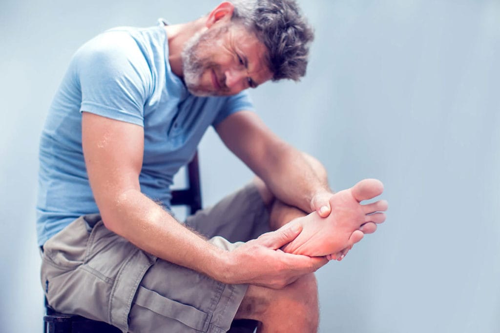
Baxter’s nerve entrapment occurs when a small nerve (known as Baxter’s nerve or, more specifically, the first branch of the lateral plantar nerve) becomes pinched (impinged) between two muscles of the inner foot. This small nerve is located on the inner aspect of the heel and when it becomes impinged it causes a sharp, burning pain, often associated with numbness and pins and needles around the heel. Baxter’s nerve entrapment can occur after injuring your ankle or heel but more commonly it develops slowly without injury and is not gender or age specific.
What are the symptoms of Baxter’s nerve entrapment?
The symptoms of Baxter’s nerve entrapment often include:
- A sharp/burning pain around the inner aspect of the heel.
- Pins and needles around the inner aspect or under the heel, especially when the nerve is knocked or tapped.
- Pain when you touch the inside of the heel.
- Pain with walking and placing your foot on the floor after a period of rest (getting out of bed in the morning).
What other conditions can present like Baxter’s nerve entrapment?
Baxter’s nerve entrapment vs plantar fasciitis
Baxter’s nerve entrapment can present very similarly to plantar fasciitis. Both normally start gradually without trauma or injury. Both give pain around the inner aspect of the heel and both affect men and women of any age. It can be very difficult to distinguish between these two conditions.
The location of symptoms and type of pain can have a subtle different. Plantar fascia pain is predominantly felt under the heel itself, whereas Baxter’s nerve entrapment is felt more on the heel and the medial arch of the foot and can also include a sensation of numbness or pins and needles.
To ensure a correct diagnosis and therefore the most effective treatment, a diagnostic ultrasound scan is essential.
What is the Baxter’s nerve?
The Baxter’s nerve, also known the first branch of the lateral plantar nerve, is a small nerve (under 1mm in diameter) running along the inside of the heel. It is an uncommon cause of heel pain, but one that should be considered when pain is not improving, particularly if you have been diagnosed with plantarfasciitis. This tiny nerve is responsible for providing muscular control to three small muscles of the foot, quadratus plantae, abductor digiti minimi and the flexor digitorum brevis. It also provides sensation to the heel bone (calcaneus) (Presley et al., 2013).

The Baxter’s nerve is a branch of a larger nerve, called the tibial nerve. The tibial nerve provides the calf muscles with both muscle control and sensation.
What is a neuropathy?
A neuropathy is an irritation of a nerve. Neuropathies can occur all over the body and often present as;
- Weakness
- Numbness
- Tingling (pins and needles)
- Burning pain
- Shooting pain
- Loss of balance
Neuropathy can be a result of a variety of different causes such as;
- Trauma - for example following an ankle sprain
- Nerve entrapment - the nerve can become pinched by other surrounding structures such as tight muscles
- Infection
- Diabetes
- Viruses such as shingles
- Some medications can also cause neuropathy
What are the symptoms of a Baxter’s nerve neuropathy?
Symptoms of a Baxter’s neuropathy include;
- Sharp or burning pain that runs along the inner aspect of the heel bone (calcaneus) and into the arch of the foot
- Tenderness of the nerve on the inside of the heel
- Pins and needles when the nerve is tapped. ‘Tapping’ the nerve to reproduce the symptoms is called Tinel’s Sign. This will be carried out by your physiotherapist.
The symptoms can be very similar to those experienced with plantarfasciitis (the most common cause of heel pain).
What causes Baxter’s neuropathy?
A Baxter’s neuropathy is predominantly a nerve entrapment issue. It occurs when the Baxter’s nerve becomes pinched or trapped as it passes under the heel (see below) through two muscles of the inner foot, the quadratus plantae and the abductor hallucis.

How do we diagnose Baxter’s nerve neuropathy?

A Baxter’s neuropathy is an uncommon cause of heel pain. It has been reported to be responsible for up to 20% of all chronic heel pain cases (Presley et al., 2013). It is often very difficult to gain an accurate diagnosis and is regularly mis-diagnosed as chronic plantarfasciitis. A delay in diagnosis can result in prolonged periods of ineffective treatment, frustration and continued pain.
For an effective treatment plan to be developed an accurate diagnosis is vital. Diagnosis includes an in-depth clinical interview, clinical testing and a diagnostic ultrasound scan. Your first appointment at Complete will include:
Clinical interview
The initial part of your assessment involves a clinical interview. Your clinician will carefully listen to your injury history and will ask you specific questions such as, how your pain started, have you had any previous treatment, what have you found helpful and what do you find particularly difficult. A full medical history is also required at this point. This helps to rule out the presence of other medical conditions that may be responsible for your condition with.
Clinical testing
After the interview you will be asked to perform a series of physical tasks. Your clinician will also feel (palpate) your foot, ankle and lower leg. Some of these tasks may be painful. The presence of pain with specific tasks can be a helpful indicator, leading to an informed diagnosis. Your clinician will also palpate the nerve specifically to determine if it causes any of your pain/symptoms.
Diagnostic ultrasound
A clinical assessment alone is not be able to formally confirm the presence of a Baxter’s neuropathy. Confirmation of a Baxter’s neuropathy can only be achieved with medical imaging techniques such as diagnostic ultrasound. Diagnostic ultrasound has been proven to be highly effective at visualising the Baxter’s nerve (Presley et al., 2013). On occasion an ultrasound guided injection local anaesthetic nerve block is required to confirm the diagnosis. This involves a small amount of local anaesthetic being injected around the nerve using ultrasound guidance to visualise the nerve and guide the procedure.
Your initial assessment at Complete will consist of both a clinical assessment and a diagnostic ultrasound scan. If you would like to book an appointment or would like more information before booking, please call 020 7482 3875 or email info@complete-physio.co.uk
How do you treat Baxter’s nerve neuropathy?
Initial treatment is often conservative, consisting of a physiotherapist led rehabilitation programme combined with manual therapy (hands on treatment) techniques. Physiotherapy treatment will include:
- Pain management advice.
- Techniques to reduce the irritation on the nerve specifically
- An individualised strengthening routine.
- Specific stretches.
- Soft tissue massage techniques.
- Acupuncture.
- Taping.
- Orthotics (inner soles for your shoes).
- Advice about how to return to exercise.
What if conservative treatment doesn’t work?
Ultrasound guided injections
A Baxter’s neuropathy is difficult to treat and does not always resolve with conservative treatment options alone. If this is the case for you then you may like to consider an ultrasound guided steroid injection. Ultrasound guided steroid injections have been proven to be highly accurate at targeting the Baxter’s nerve (Presley et al., 2013). Due to the size and depth of the Baxter’s nerve and its close proximity to a number of arteries that supply the inner heel, non-guided (blind) steroid injections are not recommended. To ensure a safe and effective injection the needle needs to be guided directly to the nerve. This can only be achieved by visualising the injection, in real time, using an ultrasound scanner.
During a guided injection a combination of a steroid (anti-inflammatory medication) and a local anaesthetic (short-term numbing agent) is introduced which bathes the nerve. This reduces the pain associated with the by reducing the inflammation.
An ultrasound guided steroid injection is very effective at reducing your pain and inflammation, however, should not be seen as a stand-alone treatment. It is vitally important that after your injection you embark on a physiotherapy program to ensure the pain does not return. Physiotherapy aims to strengthen your foot and calf musculature to improve the biomechanics of your foot particularly your foot arches.
Complete clinicians are fully qualified sonographers, independent medical prescribers and injection therapists. If an ultrasound guided steroid injection is clinically appropriate and you wish to have one your clinician will be able to prescribe the most effective medication and perform the procedure during the same appointment. The unique, one stop service means you do not need a referral from your G.P. You can simply refer yourself directly into one of our clinics.
If you would like to book an appointment or would like more information before booking, please call 020 7482 3875 or email info@complete-physio.co.uk
Other foot and ankle conditions:
- Ankle Osteoarthritis (OA)
- 1stMTP
- Chronic ankle sprain
- Morton’s’ neuroma
- Plantar Fasciitis
- Sinus Tarsi Syndrome
- Midfoot Osteoarthritis (OA)
- Calf Tear
- Gout
- Retrocalcaneal and pre-Achilles bursitis
- Mid-potion Achilles tendinopathy