Iliopsoas Tendinopathy/Bursitis
AnatomyCausesDiagnosisTreatmentInjections
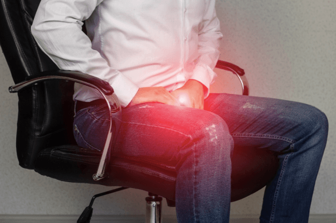
What is iliopsoas tendinopathy/bursitis?
The iliopsoas tendon is an important tendon that sits in front of the hip joint. It is the attachment for two of the main hip flexor muscles (responsible for raising your knee during activities such as putting your socks on). This tendon can become injured with repetitive movements such as running and can also become painful after a total hip replacement. When a tendon becomes irritated the healing process is compromised resulting in a thickened tendon. This is known as a tendinopathy. Occasionally the iliopsoas tendon is not injured but instead a small fluid-filled sac (bursa) becomes inflamed. This is known as bursitis. A bursitis can be very painful and present with symptoms mirroring a tendinopathy, or can even occur simultaneously.
Iliopsoas tendinopathy and/or a bursitis responds well to an ultrasound guided steroid injection if your pain is not improving or getting worse. After the injection we would recommend a course of physiotherapy to prevent the pain from returning, restore full movement and strength in the hip and get you back to full function.
What are the symptoms of iliopsoas tendinopathy/bursitis?
The symptoms of Iliopsoas tendinopathy/bursitis are:
- A deep aching pain felt in the front of your hip (in the crease of your hip).
- A clicking or clunking sensation or sound when flexing your hip (lifting your knee up).
- Sharp pain with sitting and standing, especially after prolonging sitting.
- Pain with walking, running and sport especially involving kicking and jumping.
- A feeling of hip weakness.
What other conditions present like iliopsoas tendinopathy/bursitis?
Iliopsoas tendinopathy/bursitis vs Greater trochanteric pain syndrome
Both iliopsoas tendinopathy/bursitis and greater trochanteric pain syndrome are caused by either a tendinopathy or a bursitis and can affect people of any age. Furthermore, both conditions can become inflamed with exercise or general movement. However, the location of symptoms is very different. Iliopsoas tendinopathy/bursitis can be felt at the front of your hip/groin as opposed to greater trochanteric pain syndrome which relates to pain on the outside of your hip.
Anatomy of the hip flexors
The iliopsoas tendon is the combined tendon of the two main hip flexor muscles of the hip joint; the psoas and the iliacus (see below). The psoas muscle originates from the anterior (front) aspect of all of the lumbar spine vertebrae. It passes downwards infront of the hip joint as a tendon which then terminates on the lesser tuberosity (small bony protuberance on the upper inner surface of the femur) of the femur (long bone of the thigh). The iliacus originates from the inside of the ilium (pelvis bone) and combines with the psoas tendon (forming the iliopsoas tendon) infront of the hip joint to attach onto the common insertion, the lesser tuberosity.

The iliopsoas muscle has two major functions:
- It aids in providing stability between the lumbar segments of the spine and is an important deep core stabiliser for the lower back and hip
- It provides stability and is involved in moving your knee up towards your chest (hip flexion)
This tendon can be prone to injury as it passes the front of the hip joint and is therefore a common cause of pain.
There are two main categories of patients who commonly present with a iliopsoas (hip flexor) pathology. These are:
- Runners - this is associated to repetitive movement of the iliopsoas tendon over the front of the hip during exercise such as running. The tendon and the bursa (a small sac surrounding the tendon) can become inflamed. This process can be exacerbated by poor biomechanics and tissue overload (‘doing too much too soon’) and can be due to both muscle weakness and tightness in the surrounding hip musculature.

- Post total hip replacement (see below) patients - the iliopsoas tendon and the bursa can become swollen and/or the inflamed (discussed in more detail below) as it travels over the metal implant. This reportedly presents in 4.3% of patients undergoing a total hip replacement (Nunley et al., 2009).

Iliopsoas tendinopathy/bursitis has also been associated with rheumatoid arthritis and hip osteoarthritis (Garala et al., 2014).
How does the iliopsoas major tendon and bursa become injured?
Iliopsoas tendinopathy/bursitis is predominantly an overuse injury. As the iliopsoas tendon passes the front of the hip joint it can come impinged (pinched). Repetitive compression or pinching of the tendon causes an inflammatory reaction to occur. Inflammation of the tendon is called tendinitis. Tendinitis is often short lived and self-limiting, usually resolving within a few weeks of rest. When the iliopsoas tendon is subjected to multiple bouts of tendinitis with insufficient recovery, the tendon structure becomes weaker. Driven by a maladapted healing process the tendon also becomes thickened and compromised. This process is commonly called tendinopathy. It is not uncommon in this condition to experience associated ‘snapping’ or clicking at the front of the hip. Sometimes the ‘clicking’ causes pain but it can often be asymptomatic (painfree).
Surrounding the iliopsoas tendon at the level of hip joint is a small sac or cushion called a bursa. Bursa contain a naturally occurring lubricant known as synovial fluid. There are over 150 bursae located throughout the body, found in ‘anatomical friction hotspots’. These protective structures are perfectly designed to allow frictionless gliding between structures during movement. However, if they are exposed to repetitive movements (as in running) or trauma (in this case total hip surgery) the bursa can become inflamed. Bursal inflammation is known as bursitis. Iliopsoas bursitis is relatively rare and can be associated with an underlying problem with the hip joint such as such as osteoarthritis. It is therefore critical that a full and accurate assessment of the hip be completed when diagnosing iliopsoas tendinopathy/bursitis.
How do you know if you have iliopsoas tendinopathy/bursitis?
Both iliopsoas tendinopathy and bursitis can be very painful and limit your activities of daily living.
Common symptoms of iliopsoas tendinopathy/bursitis include:
- Pain - as in a deep aching pain located at the anterior aspect of the hip.
- A ‘clunking’ or ‘clicking’ sensation deep within the hip often experienced during both hip flexion (raising the knee to the chest) and/or hip extension
- Pain when standing from a seated position
- Pain during or after prolonged sitting
- Pain with walking and exercise, especially running
- Sensation of weakness in the muscles surrounding the hip and thigh
Prior to receiving treatment for your symptoms is essential that an accurate diagnosis is made to ensure the correct treatment is recommended. It is therefore important to seek professional advice.
A diagnosis of iliopsoas tendinopathy/bursitis requires a combination of clinical physical assessment and diagnostic ultrasound imaging.
Clinical physical assessment can be undertaken by one of our highly experienced clinicians at Complete and often involves:
- An in-depth discussion surrounding the history of your condition. Direct questions often include how your pain started, what aggravates the pain and what eases your symptoms. You will also be asked questions regarding general medical health to assess for systemic causes of your pain, such as rheumatoid arthritis. If your clinician suspects systemic cause of your symptoms, you may be referred for blood tests to help formulate a diagnosis.
- Hip joint range of movement.
- Hip muscle strength testing.
- Lower limb flexibility.
- Palpation (feeling) of the hip and lower back.
- Functional testing involving squatting technique and walking and running assessment (if you are a runner).
The clinical assessment process enables the clinician to formulate a working hypothesis. It is also able to assess for biomechanical insufficiencies which may be aggravating and/or the underlying cause of your pain.

Clinical assessment alone is unable to diagnose iliopsoas tendinopathy/bursitis. For a definitive diagnosis, diagnostic imaging is required.
Diagnostic imaging is also able to differentiate other causes of hip pain which may be masquerading as iliopsoas tendinopathy/bursitis or may be occurring as well as a tendon issue.
- Osteoarthritis of the hip
- Femoral acetabular impingement of the hip (FIA). FIA is a common condition where the ball and socket joint of the hip develops a small bony prominence. A bony prominence can occur on the acetabulum (socket of the hip) and is called a pincer lesion or on the neck of the femur, called a cam lesion. It occurs in up to 30% of the population. During movement, the bony prominence catches resulting in an impingement within the joint.
- Label hip tears. The labrum is a crescent-shaped, fibrocartilaginous rim that surrounds the socket of the hip (acetabulum). This structure increases the depth of the socket, increasing the joints stability. It can get torn due to injury or secondary to femoral acetabular impingent (FAI) or osteoarthritis of the hip.
- Greater trochanteric pain syndrome (GTPS). This causes pain on the outside of the hip and is secondary to gluteus tendinopathy and/or an associated bursitis (known as trochanteric bursitis).
X-ray

An X-ray cannot diagnose iliopsoas tendinopathy or bursitis. X-ray imaging (as seen above) is a highly effective diagnostic tool for diagnosing bone and joint pathology and is routinely requested to assess for osteoarthritis. This gold standard imaging technique has been used for many decades and is capable of not only diagnosing joint pathology but also assessing the severity of the condition. If your clinician suspects osteoarthritic change within the hip joint they may refer you for an x-ray.
Magnetic resonance imaging (MRI)

MRI imaging is capable of assessing both joint and soft tissue pathologies and is often requested for assessing iliopsoas tendinopathy/bursitis, femoral acetabular impingement (FAI) and/or labral tears. An MRI can take between 30 minutes and an hour and involves you lying very still within the metal tube. The MRI machine takes multiple images, from front to back, of your hip. These slices provide a highly detailed 3D image of your hip structure.
Diagnostic ultrasound imaging
At Complete we carry out a diagnostic ultrasound at every appointment, at no extra charge. Diagnostic musculoskeletal ultrasound imaging is very effective to assess tendon and bursal pathology around the hip. Diagnostic ultrasound is the only imaging modality which allows us to assess the anatomical structures as you move. We call this a dynamic ultrasound scan. This is excellent to assess for ‘clicking’ and ‘clunking’ of the hip.
It is, therefore, a highly effective imaging tool to assess for both the iliopsoas tendinopathy and bursitis during movement to assess for pain and clicking associated with this pathology (Garala et al., 2014. Balus et al., 2014). At complete our team of clinicians are qualified as both physiotherapists and musculoskeletal sonographers. During your initial assessment, your clinician will conduct a thorough clinical assessment as well as a diagnostic scan.
If you would like more information or would like to book an appointment please contact us on 0207 4823875 or email injections@complete-physio.co.uk.
How do we treat Iliopsoas tendinopathy/bursitis?
The vast majority of patients respond very well to conservative management. Physiotherapy forms the foundation of your management for iliopsoas tendinopathy/bursitis. Physiotherapy often involves:
- An individualized progressive rehabilitation program involving stretching and strengthening exercises for the hip musculature.
- Advice on activity modification to reduce aggravating factors for your hip
- Biomechanical re-education. This may include teaching you good form during squatting or lunging techniques
- In some cases, soft tissue release techniques are used to reduce pain and increase flexibility around your hip. You may also be taught some self-massage techniques using a foam roller.

What if conservative treatment does not work?
If conservative management has not been effective at reducing your symptoms and you are still experiencing pain remains injection therapy may be appropriate for you.
There are two types of patients suffering from psoas tendinopathy/bursitis who commonly require an injection:
- Runners, triathletes or ironmen often experience pain and symptoms due to repetitive friction of the iliopsoas tendon and bursa over the front of the hip.
- After a total hip replacement. Patients can be susceptible to iliopsoas tendinopathy and/or bursitis after a total hip replacement. This often occurs due to friction of the bursa and tendon over the new metalwork. Patients experiencing these symptoms can respond very well to injection therapy. Injection therapy for this patient group is closely discussed with your surgeon prior to completing an injection.
Injection therapy is not a stand-alone treatment. It provides you with a pain-free ‘window of opportunity’ to rehabilitate your hip effectively. Complete recommend after undertaking an injection that a course of physiotherapy starts within two weeks to ensure best results are achieved.
Injection therapy is an excellent technique for reducing pain and inflammation and is particularly effective if you are suffering from one or more of the following:
- Pain is waking you at night or stopping you from sleeping.
- Pain is limiting you from completing activities of daily living or sporting/leisure activities.
- Pain is restricting you from completing a physiotherapy rehabilitation program.
Due to the depth of the iliopsoas tendon and bursa, as well as the close proximity of the arteries, veins and nerves that supply the leg it is essential that an injection is ultrasound-guided. Ultrasound-guided injections have been shown to be highly accurate at delivering the medication to the target tissue. During this technique, real-time ultrasound imaging is used to guide the needle tip directly to the source of the symptoms. Due to this highly accurate technique, post-injection complications can be minimised. Research has shown ultrasound-guided injections to be more efficacious and has fewer complications than Landmark guided injections.
Ultrasound-guided steroid injection
Ultrasound-guided corticosteroid injection is an effective treatment option for persistent pain associated with iliopsoas tendinopathy/bursitis (Garala et al., 2014 and Nunlet et al., 2009). During this guided technique a small amount of corticosteroid (powerful anti-inflammatory medication routinely used within injection therapy) is combined with a short-acting local anaesthetic. The consensus within the research states on average 10 to 12 weeks for a significant reduction in pain after receiving an ultrasound-guided corticosteroid injection. This window of opportunity will enable you to maximise your rehabilitation.
At Complete all our clinicians are fully qualified musculoskeletal sonographers and independent prescribers. Your appointment includes a full and accurate diagnosis including a diagnostic ultrasound scan and if required an ultrasound-guided injection.
If you would like more information or would like to book an appointment please contact us on 0207 4823875 or email injections@complete-physio.co.uk.
Other hip conditions
Greater trochanteric pain syndrome
Hip osteoarthritis
Proximal hamstring tendinopathy
Femoroacetabular hip impingement (FAI)
References
Garala, K., Prasad, V., Jeyapalan, K. and Power, R.A., 2014. Medium-term and long-term outcomes of interventions for primary psoas tendinopathy. Clinical Journal of Sport Medicine, 24(3), pp.205-210.